- Clinical
- Imaging & Planning
- Ancillary Services
- Positioning and Immobilisation
- Treatment Delivery
- Veterinary
- MRI Solutions
- SABR Solutions
- Physics
- QA
- Imaging QA
- Patient QA
- Machine QA
- Encephalon 3D phantom
- myQA® Daily
- myQA® Machines
- myQA StarTrack³
- MatriXX Resolution
- Prime phantom
- QUASAR™ Heavy Duty Respiratory Motion Platform
- QUASAR™ MLC
- QUASAR™ MRgRT Insight Phantom
- QUASAR™ Motion MR Platform
- QUASAR™ Penta-Guide Phantom
- QUASAR™ Respiratory Motion Phantom pRESP 2.0
- SBRT phantom
- Spine phantom
- StarTrack Detector
- Dosimetry
- Proton QA & Dosimetry
- Software
- MRI Physics Solutions
- QA
- SGRT
- Medical Imaging
- Exterior and Interior Design
- Manufactured by
- Support & Finance
- News
- About
- Contact
QUASAR™ MRID3D
A novel method for quantifying 3D geometric distortion in MRI

Overview
QUASAR™ MRID3D is a lighter, larger and more efficient way to quantify MRI geometric distortion in 3D! Analysis is performed on images for MRgRT, simulation and diagnostic imaging applications.
Unlike other distortion analysis products, which only sample a few imaging planes, the QUASAR™ MRID3D Geometric Distortion Analysis System analyses the entire 3D volume in a single series acquisition.
The system uses a novel method based on 3D harmonic analysis of distortion by analysing the Boundary Distortion Vector Field derived from the location of known control points on the surface of the phantom. 3D Laplace Partial Differential Equations are used to calculate the entire 3D Distortion Vector Field.
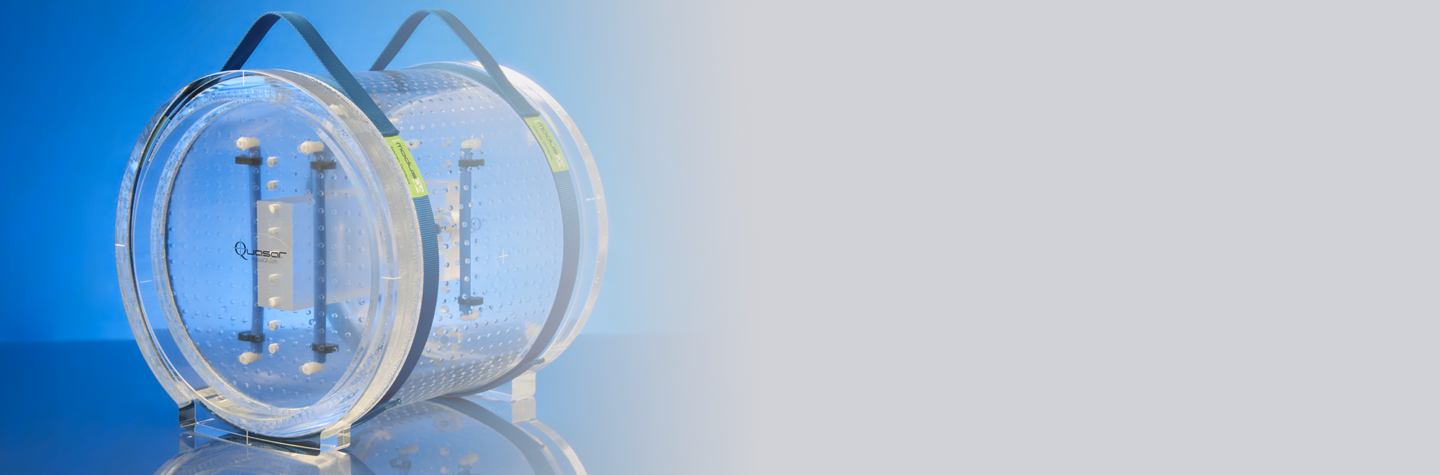
The large, acrylic, and hollow boundary phantom features integrated feet and handles for fast and easy setup in MRI scanners and includes image analysis software with a built-in 3D DICOM Viewer and Region of Interest (ROI) selector permitting user analysis of smaller custom volumes.
MRID3D Phantom video
Benefits
- Follows NEMA/MITA MS-12 →recommendations and IEC 62464-1 standards for highest accuracy and precision
- Acquire T1 weighted 3D isotropic GRE scans at 3T under 5 minutes or at 1.5T as quick as 10 minutes
- Uses a novel method for quantifying 3D geometric distortion in MRI based on 3D harmonic analysis
- Features an XYZ orientation and scale cuboid containing fiducials channels to easily visualize the phantom isocentre
- Contains 1,496 precisely machined fiducial markers uniformly distributed around the phantom surface boundary
- Plus 6 precisely machined reference markers to detect image position when distortion is high or correction is off
- Enables users to easily visualize analysis results and update visually informative charts, graphics and plots in real-time
- Permits users to update statistical analysis in real-time within a user defined 3D region of interest
- User selects 2 identical MR scans with opposite frequency encode polarities to compare B0 vs Gradient distortion
- Software automatically locates fiducials, compares it against the truth and calculates the geometric distortion for QA analysis
- Track results over time to identify patterns, prevent potential errors and make adjustments to improve patient care
- Capable of exporting the entire 3D Distortion Vector Field (DVF) data into Microsoft Excel (.CSV) format
- Save distortion measurement results in PDF format to share with physicist, MRI technician and radiation therapists
- Designed for multiple QA applications including MRI-guided radiation therapy, MR simulation and diagnostic imaging
- 37cm x 32cm (W x L) phantom enables users to accurately simulate and QA treatment plans for a wide range of patient sizes
- Phantom weighs 21 kg for easy and fast setup compared to similar sized water-based phantoms that weigh 41 kg or more
- The removable end plate, XYZ cuboid and fiducial markers are delivered pre-filled with mineral oil for maintenance free use
- The system never has to be drained or refilled and comes complete with analysis software and instructions for safe handling
Key Features
- Phantom Design
- Fast Acquisition
- Patented Technology
- Isocentre Detection
- Fiducial Markers
- Reference Markers
- Feature-rich Graphics
- Built-in 3D DICOM Viewer
- Reverse Polarity Analysis
- Automated Analysis
- Trend Analysis
- Export Results
- Detailed Reports
- Versatile Usage
- Large 3D Volume
- Hollow Phantom
- Waterless Phantom
- Complete System
A better phantom measurement technique for modern MRgRT applications.
The QUASAR™ MRID3D Phantom contains an array of 1,502 precisely machined mineral oil filled fiducial markers for accurate and robust position detection. The 3D spatial position of each control point, defined as the centroid of the closed end of each fiducial, is consistently fabricated to within 0.1 mm relative to the truth template derived from CAD data.
The fiducial markers with 18 mm uniform spacing conform to the NEMA MS-12 standard for precision, quantification and mapping of geometric distortion in Magnetic Resonance Imaging.
Conformance to recommendations on design and data acquisition by The Medical Imaging & Technology Alliance (MITA), a division of the National Electrical Manufacturers Association (NEMA), enables the QUASAR™ MRID3DPhantom to accurately measure MR image distortion at low and high field strengths, even under conditions of large distortion such as low bandwidth acquisitions with vendor distortion correction turned off.
Designed for geometric dimensional stability.
The temperature compensating oil expansion reservoirs maintain geometric dimensional stability of the phantom when temperature variations are experienced in use. This eliminates a potential source of measurement error commonly found in large conventional grid phantoms, and ensures safe, leak-proof shipping.
An XYZ orientation and scale cuboid insert, located at the centre of the phantom, is used to register a calibrated frame of reference and to facilitate auto pre-scanning 3 plane MR Image localisers.
Maintenance free and easy.
The physical dimensions of the phantom are 39.4 cm diameter by 39.1 cm long – with a hollow internal volume of ~25 litres, effectively removing 25 kg of weight relative to conventional solid fluid filled grid phantoms. At 21 kg the maintenance free phantom from Modus QA is half the weight of conventional solid fluid filled grid phantoms for lighter, convenient one-person setup.
The phantom comes pre-filled with high T1 weighted contrast mineral oil for faster 3D scanning and dielectric-resonance-free use at high 3T fields. The mineral oil is susceptibility matched to acrylic to render susceptibility artefact negligible around the control points.
Software that meets the demand for accuracy, speed, and efficiency.
The QUASAR™ MRID3D software application is designed with specific needs of MR medical physicists and clinical workflow in mind. Accuracy, speed, and efficiency of results, were primary considerations during the design process.
The application is optimized to run locally without any load or latency issues, which translates into less time waiting for cloud-based applications to generate 3D distortion vector fields or for a spinning wheel to stop spinning.
Algorithms that calculate the entire 3D Distortion Vector Field, automatically.
The robust software automatically identifies the location of the isocentre and control points in the acquired MR images of the phantom and uses a fully automated process to compare them against the truth to calculate the geometric distortion.
The geometric moment of distortion truth has arrived.
Sophisticated imaging processing techniques are automatically enabled to measure the distance between the locations of the fiducial markers in the distorted MR image and their known positions defined by the geometry of the phantom.
The characterisation and measurement results of the geometric distortion in MR images are displayed in 3-dimensions with unprecedented details and accuracy for QA analysis.


Found what you're looking for or need to discuss your requirements?
Call us today on +44 (0)1743 462694 or email us here
Access your product documents and software downloads via OSL Portal